
Impacted Wisdom Tooth Surgery: When to Remove It and What to Expect
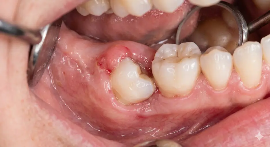
Wisdom teeth, or third molars, are the last teeth to appear, usually between the late teens and mid twenties. Many jaws do not have enough room for them, so they get stuck against the gum, bone or the tooth in front. A tooth that cannot fully come through is called impacted.
Not every impacted tooth must be removed, but those that cause problems are best treated early, before infection or damage to neighbouring teeth develops.
Types of impaction
- Mesioangular: tilted forward toward the second molar, the most common pattern
- Vertical: upright but trapped under gum or bone
- Horizontal: lying sideways against the second molar
- Distoangular: tilted toward the back of the mouth
The pattern, together with the depth of the tooth and the position of the nerve, decides how difficult surgery will be.
Why impacted wisdom teeth cause problems
- Pericoronitis: repeated painful gum infection around a partly erupted tooth
- Decay: food and plaque trap between the wisdom tooth and the second molar, damaging both
- Damage to the neighbouring tooth: pressure or decay on the second molar's root
- Cysts: fluid-filled sacs forming around a buried tooth, rarely a tumour
- Pain, swelling, bad taste and limited mouth opening
When removal is recommended
Removal is advised when there is recurring infection, decay, cysts, damage to the neighbouring tooth, gum disease, or when the tooth is needed to be removed before orthodontic or other treatment. For a wisdom tooth with no symptoms and a healthy position, observation with regular X-rays may be sensible, and your dentist should explain both options.
Planning the surgery with 3D imaging
A panoramic X-ray is usually the starting point. When the roots lie close to the nerve in the lower jaw or to the sinus in the upper jaw, a cone beam CT gives a three-dimensional view and helps plan the safest approach. Read how this works in our article on 3D imaging in dentistry.